Materials Science and Non-Biological Applications
The Transition from Biological to Materials Science
Historically, Serial Block-Face Scanning Electron Microscopy (SBF-SEM) was conceived and optimized for high-resolution, three-dimensional (3D) reconstructions of soft biological tissues [271]. In these early life-science applications, the technique revolutionized the mapping of neuronal networks and cellular ultrastructure by automating the tedious process of serial sectioning [271]. However, the inherent capability of SBF-SEM to generate perfectly aligned tomographic datasets with nanoscale lateral and axial resolution has driven a profound expansion of its utility into materials science and hard condensed matter [12, 56, 271] (Figures 57–58).
The transition to materials science applications was notably spearheaded by Zankel et al. in 2009, demonstrating that the in-chamber ultramicrotomy previously reserved for lipid-rich cells could be successfully applied to synthetic structures [271, 272]. Today, SBF-SEM is increasingly employed to investigate a vast array of non-biological media, spanning functional polymer blends, organic coatings, structural alloys, sintered metals, cementitious materials, and complex geological samples [12, 56, 271]. For materials scientists, the technique offers an unprecedented capacity to quantify phase distributions, porosity, and internal microstructural defects over large analytical volumes, effectively bridging the spatial resolution gap between highly localized Transmission Electron Microscopy (TEM) and lower-resolution X-ray Computed Tomography (XRT) [56, 272] (Figure 59).
Polymers, Paints, and Complex Composites
The evaluation of complex polymer networks and composite materials has greatly benefited from the exceptional Z-axis resolution provided by SBF-SEM [12]. In the realm of separation science and chromatography, characterizing the internal geometry of polymer-based monolithic stationary phases has historically been challenging due to the soft matter nature of the materials [273]. SBF-SEM has been successfully utilized to reconstruct the 3D morphology of hyper-cross-linked poly(styrene-divinylbenzene) monoliths [273]. To achieve sufficient backscattered electron (BSE) contrast and mechanical stability during diamond knife microtomy, the polymer skeleton was heavily stained with tetraphenyllead, and the internal void spaces were infiltrated and embedded with epoxy resin [273]. The resulting volumetric data allowed for sophisticated chord length distribution analyses, which identified two distinct macropore types with characteristic lengths of 7.32 µm and 0.73 µm [273]. Furthermore, quantitative mapping revealed a macroporosity averaging 77% that systematically increased from the capillary wall toward the center of the monolith, directly informing the hydrodynamic properties and synthesis parameters of the stationary phase [273] (Figure 60).
Beyond pure polymer frameworks, SBF-SEM has elucidated the internal architectures of highly heterogeneous, multi-phase industrial coatings and paints [78, 271]. In barrier marine coatings designed for extreme environments, the percolation properties and spatial arrangements of functional fillers are critical to anti-corrosive performance [78]. SBF-SEM has provided clear, contrast-reversed BSE datasets distinguishing aluminum flakes, talc fragments, iron oxide nanoparticles, and the continuous epoxy resin matrix [78]. Notably, the 3D segmentations revealed a parallel, stratified arrangement of the aluminum flakes that was entirely indiscernible in conventional 2D cross-sections [78]. This spatial alignment fundamentally dictates the barrier efficacy and solvent resistance of the coating [78]. Similarly, SBF-SEM has been deployed to examine waterborne paints containing titanium dioxide (TiO2) pigments within an acrylic resin matrix [275]. By tracking the 3D distribution of the high-atomic-mass TiO2 particles, the surrounding acrylic binder, and internal voids, researchers perform rigorous threshold segmentations to analyze the micro-environmental encapsulation of pigments [275]. This capacity to directly correlate formulation parameters to final optical and protective properties is highly sought after in the coatings industry [275].
The technique has also been adapted to study electrical degradation in dielectric polymers, notably through the visualization of "electrical trees" [15]. Electrical trees are complex, branching micro-channels of dielectric breakdown that propagate through insulating polymers under sustained high electrical stress [15]. Using low-vacuum SBF-SEM, researchers can reconstruct 3D models of these fractal-like void networks, bypassing the limitations of traditional targeted sectioning which only captures isolated 2D branches of the breakdown path [15].
Metallic Alloys and Sintered Nanoporous Structures
Despite the formidable physical challenges associated with the ultramicrotomy of dense metals, SBF-SEM has proven to be an invaluable tool for characterizing the internal structures of hard metallic systems [12, 34, 41, 56, 271]. In lightweight structural materials, such as aluminum-silicon casting alloys, 3D imaging has been utilized to map the spatial morphology and phase distribution of specific intermetallic inclusions [41]. The highly aligned, sequential SBF-SEM slices enable the quantitative visualization of crack propagation paths and growth relationships within the alloy matrix, offering insights into mechanical failure mechanisms [8, 107].
Furthermore, SBF-SEM provides critical empirical data required for the micromechanical modeling of highly porous metals [65, 272]. For example, the technique has been utilized to characterize nanoporous aluminum and nanoporous gold fabricated via vapor-phase and chemical dealloying processes [65, 66]. Due to the large field of view combined with high voxel resolution (e.g., ), SBF-SEM can capture tens of cubic microns of the highly complex, continuous ligament-and-pore networks characteristic of these materials [65, 66]. To analyze these datasets, digitized structural arrays are converted into voxel indicator functions that mathematically distinguish the solid phase from the void phase [65]. This enables the precise calculation of normalized centers of mass and exact solid volume fractions across hundreds of sequential slices [65]. These image-based structural arrays serve as direct inputs for Finite Element (FE) simulations [65]. By translating SBF-SEM data into computational meshes, researchers can calculate macroscopic mechanical properties, such as Young's modulus, moving beyond idealized mathematical representations (like spinodal or gyroidal microgeometries) to accurately reflect the true morphological heterogeneity of the fabricated metal [65].
Similar imaging-to-simulation workflows have revolutionized the study of sintered micro-silver pastes, which are increasingly utilized as lead-free die-bonding joints in microelectronics [272]. SBF-SEM facilitates the high-resolution segmentation of pore fractions, shapes, and spatial distributions within the sintered silver network, both immediately after processing and following extensive thermal aging [272]. By generating stereolithography surfaces at the pore/matrix interfaces, researchers import these meshes into finite element solvers to simulate the elastic behavior of the porous solid under stress [272]. The Young's modulus derived from these authentic 3D SBF-SEM geometries closely matches experimental values obtained via dynamic resonance methods [272]. These integrated studies have confirmed that thermal aging at 125 °C for 1500 hours does not alter the density or Young's modulus of the silver joint, demonstrating that the elastic modulus of the sintered material is fundamentally a function of its pore fraction [272].
Cementitious Materials: Tricalcium Silicate Hydration
Arguably one of the most extensive and impactful non-biological applications of SBF-SEM to date is the microstructural analysis of cementitious materials, specifically the hydration of tricalcium silicate (3CaO·SiO2, or C3S) [41, 271]. C3S constitutes 50–70% of the mass of ordinary Portland cement and dominates early-stage strength development and normal setting behavior [41, 271]. Understanding its complex 3D microstructural evolution is paramount for optimizing concrete durability; yet, traditional 2D SEM provides limited spatial context [41, 271], while XRT lacks the nanoscale resolution required to observe fine gel and capillary pores [271]. SBF-SEM perfectly bridges this analytical gap [41, 271].
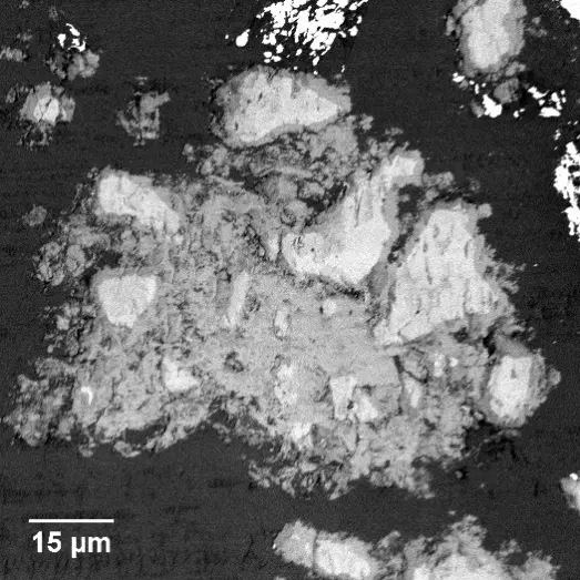
The characterization of raw, unhydrated C3S powders embedded in epoxy resin has revealed highly irregular, quasi-acicular morphologies [41]. To ensure high-quality data, samples are rigorously prepared, with C3S purities reaching 98% via the Rietveld method prior to embedding [271]. Quantitative 3D analysis from SBF-SEM established that most C3S particles exhibit length-width ratios (LWRs) between 3 and 5, rather than the ideal spherical geometries frequently assumed in classical hydration models [41]. Such shape factors directly increase the theoretical water-to-cement ratio required for complete hydration [41]. Furthermore, SBF-SEM detected a remarkably rich internal pore structure within large individual grains, discovering completely enclosed pores and open channels penetrating the entire particle [41]. This internal porosity significantly increases the effective surface area available for aqueous reactions, directly accelerating hydration kinetics [41].
Upon mixing with water, C3S reacts to form calcium silicate hydrate (C–S–H) gels and crystalline calcium hydroxide [271]. In samples hydrated for 24 hours, SBF-SEM BSE imaging clearly differentiates bright anhydrous C3S cores, grey hydration products, and dark, resin-filled capillary pores [271]. Volumetric SBF-SEM datasets have enabled the precise quantification of capillary pore networks, which ultimately govern the macroscopic permeability, shrinkage, and mechanical strength of concrete [271]. In one comprehensive reconstruction, 4,800 individual pores were segmented, revealing a total porosity of approximately 9%, of which nearly half consisted of interconnected pore clusters critical for water transport [271]. The average capillary pore volume was measured at , with diameters predominantly ranging below 2 µm [271].
While the spatial resolution of SBF-SEM (typically ~50 nm in these highly insulating cementitious samples) cannot resolve the finest nanoscale gel pores (0.5 to 10 nm), the technique provides vastly superior morphological and connectivity data for capillary pores compared to bulk techniques like mercury intrusion porosimetry (MIP) [271]. MIP struggles to accurately differentiate closed versus open porosity due to "ink-bottle" effects, whereas SBF-SEM visually captures both independent and connected void networks [271]. Furthermore, 3D distance mapping quantified the distribution of these pores relative to the anhydrous C3S cores, identifying an inflection point at a distance of 860 nm [271]. This empirical finding structurally validates the well-known "inner and outer product" kinetic models of cement hydration, mapping exactly where specific hydrate phases precipitate in 3D space [271].
Geological Samples, Zeolites, and Minerals
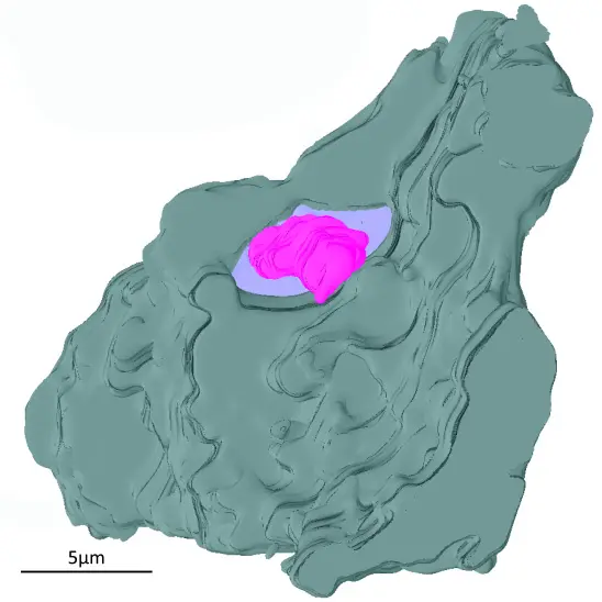
Beyond cements, SBF-SEM has also been extended to porous geological samples and complex industrial minerals [41, 274]. For example, SAPO-34 zeolite crystals—which are widely used as catalysts in the petrochemical industry—exhibit low electrical conductivity and high physical hardness, presenting significant characterization challenges [40, 271, 274]. By employing modified sample embedding techniques, researchers have successfully utilized SBF-SEM to explore the internal microstructure of SAPO-34 powders [274]. The resulting tomograms successfully mapped 3D grain structures, internal crystalline domains, and varied micro-cracking patterns hidden deep within the particles [274]. These direct, real-space 3D observations corroborate indirect findings from non-destructive synchrotron techniques like Bragg Coherent Diffraction Imaging (BCDI), definitively confirming the presence of imperfect, highly defected internal crystal domains that heavily influence the material's catalytic performance [274].
Methodological Adaptations for Hard Condensed Matter
The expansion of SBF-SEM into materials science has necessitated significant methodological innovations, primarily to address two formidable technical challenges: specimen charging and extreme diamond knife wear [14, 18, 78, 271, 274]. Unlike lipid-rich biological tissues that can be heavily osmicated with heavy metals to intrinsically increase electron yield and tissue conductivity, inorganic crystals, cement minerals, and synthetic polymers are inherently insulating and prone to severe electron accumulation [14, 271, 274]. This localized charging repels the primary electron beam and distorts the emitted BSE signal, severely degrading image resolution, causing image drift, and introducing dark-band artifacts across the dataset [14, 271].
To mitigate charging in materials science applications, conventional epoxy resins are frequently modified with highly conductive additives prior to embedding [18, 34, 38, 119, 274]. The incorporation of carbon black nanoparticles (e.g., Ketjen black) or metallic silver particles into the liquid resin matrix creates conductive pathways that effectively dissipate accumulated surface charge away from the block face during imaging [18, 34, 38, 119, 274]. Additionally, researchers typically ground the external surfaces of the polymerized resin block by coating it with conductive silver paint or by sputter-coating the initial block face with a thin layer of gold [41, 65]. When sample composition inherently precludes high-vacuum imaging despite conductive resins, environmental or variable-pressure SEM modes are utilized [12, 14, 15, 275]. By introducing controlled amounts of water vapor or nitrogen gas (typically 10 to 70 Pa) into the specimen chamber, the electron beam ionizes the gas molecules, which in turn neutralize the negative charge accumulating on the sample [12, 14, 15, 275]. This restores image fidelity and prevents structural distortion, albeit occasionally at a slight compromise to the ultimate signal-to-noise ratio and attainable voxel resolution [12, 14, 15, 275].
Finally, the sheer physical hardness of metals, ceramics, and cementitious phases exerts exceptional wear on the ultramicrotome's diamond knife [14, 18, 78]. Slicing artifacts—such as knife chatter, physical scoring of the block face, or complete sample avulsion from the resin—are constant risks when sectioning hard condensed matter [14, 18, 78]. Consequently, researchers must meticulously optimize cutting parameters for inorganic samples. This often involves reducing the slice thickness to the 15–50 nm range and substantially decreasing the traverse speed of the diamond knife to maintain a pristine block face throughout extensive automated acquisitions [41, 78, 271].
Through these continuous adaptations in embedding chemistry, charge compensation, and variable-pressure electron optics, SBF-SEM has firmly established itself as a transformative analytical tool in materials science. From unlocking the nano-mechanics of porous metals to visualizing the fundamental hydration kinetics of modern cements, the technique provides the critical third dimension required to link complex microstructural heterogeneity to macroscopic material performance.